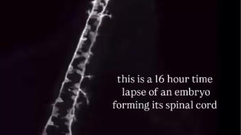
Does an often reposted viral video show a timelapse of "16 hours of spinal cord development" in a human embryo? No, that's not true: The video actually shows the development of the nervous system of a zebrafish as recorded by researchers from Wisconsin. The video is often reposted without that bit of information omitted, leading many people online to believe it shows a human embryo.
An example of the video can be seen in this post on X (archived here) published on November 8, 2025 with a caption that read:
16 hours of spinal cord development in an embryo captured in stunning timelapse
16 hours of spinal cord development in an embryo captured in stunning timelapse pic.twitter.com/dN5ThzSpZC
-- ViralRush ⚡ (@tweetciiiim) November 8, 2025
The video had a text caption that read:
this is a 16 hour time lapse of an embryo forming its spinal cord

(Image source: screenshot of ViralRush video)
However the video already appeared rotated by 90 degrees in a 2018 article (archived here) titled "Light Sheet Imaging Helps Capture Zebrafish Neural Development" where it was described as:
The footage, imaged using light sheet microscopy, is credited to Henry He, a scientist in Jan Huisken's lab at the Morgridge Institute for Research, and Liz Haynes, a postdoctoral fellow in Mary Halloran's lab at the University of Wisconsin-Madison.
...
The video shows the development of a zebrafish embryo over a period of 16 hours. In discussion with Technology Networks, Liz and Henry go into more detail on exactly what we are seeing in the footage: "The embryo being imaged is a genetically modified organism expressing green fluorescent protein in a population of its sensory neurons. During the beginning of the movie, the main focus is on two rows of neurons in the spinal cord of the embryo (we are looking at its back and side). The cell bodies of these neurons extend two axons each in the spinal cord, forming a network to talk to the brain. They then send out a third axon, called a peripheral axon, which exits the spinal cord and innervates the skin of the embryo so it can sense touch. These axons grow incredibly long distances and establish complex, beautiful architectures." By the end of the video, the tip of the embryo's tail can be seen growing back into the movie's focal plane.
The video from the article (archived here) can be viewed below:


